


Description
Quick Details
Acquire Precise Images with X Line Objectives
Bright LED Lighting Designed for Pathology and Laboratory
Bright Images in Multi‐Head Configurations
Coded units to integrate with imaging software
Packaging & Delivery
| Packaging detail:Standard export package Delivery detail:within 7-10 workdays after receipt of payment |
Specifications
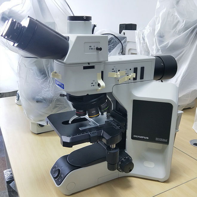
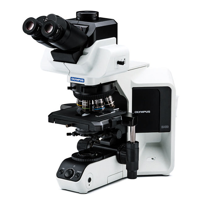
Teaching and Challenging Applications Olympus Microscope BX53
With an LED illuminator equivalent to or better than a 100 W halogen lamp, the BX53 microscope delivers brightness that’s appropriate for teaching and various contrast methods. Customize your microscope with modular units based on the observation methods you want to use. Choose from options including condensers, nosepieces, a rotating stage, objectives, and intermediate optics optimized for various observation methods, including phase contrast and fluorescence.
Acquire Precise Images with X Line Objectives
Improved flatness, numerical aperture, and chromatic aberration combine to deliver clear, high‐resolution images with excellent color reproduction. The objectives’ superior chromatic aberration management delivers better color accuracy across the entire spectrum. The elimination of violet color aberration creates clear whites and vivid pinks, improving contrast and sharpness.
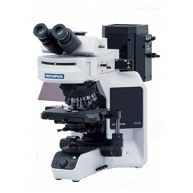
Bright LED Lighting Designed for Pathology and Laboratory
Designed with spectral characteristics that mimic halogen light sources, the BX3 series’ LED illumination enables users to clearly view the purple, cyan, and pink colors important in pathology, but typically difficult to see using LEDs. Users get the benefits of an LED, including consistent color temperatures and long use life, without the typical trade offs.
Bright Images in Multi‐Head Configurations
Multi‐head discussion systems are essential for training and education. With the BX53 microscope’s LED illumination, up to 26 participants can view clear, bright images.
Coded units to integrate with imaging software
Add an optional coded nosepiece to your BX53 microscope to automatically record and share magnification setting information for post‐imaging treatments. The metadata is automatically sent to cellSens software, helping minimize mistakes and scaling errors.